MANTIS® SPATIAL BIOLOGY PLATFORM
Multiplex ANnotated Tissue Imaging System
Spatial biology studies as a service
Human data from fluorescence-based multiplex 3D-imaging

The MANTIS® spatial biology platform is Genoskin’s unique Multiplex Imaging System that allows you to study skin biology within its spatial context in order to obtain relevant and reliable data on the response of real human skin to your vaccine, drug or cosmetic. Spatial skin biology gives a new dimension to single-cell analysis as it helps to understand skin cell organization within the epidermis, dermis and hypodermis. It also provides crucial information on cell interaction and on the simultaneous response of different cells to the compound you’re studying. Genoskin is proud to offer this unique technology as a service.
Multiplex
Mantis® is a 3D multiplex imaging platform to simultaneously visualize skin immune cell populations in one single pass.
Human
We use real human data based on the live response from our skin platforms or from the clinical samples you provide.
Skin
Our experts have 10 years of experience in using skin biology and immunology as a window to the human body.
Immunoprofiling
We provide dedicated study panels to simultaneously analyze a wide range of human immune cells and get the results you need.
Automated & simultaneous spatial skin analysis
Visualize over 12 markers in 1 acquisition
The MANTIS® spatial skin biology platform is a flexible and interactive analytical system, specifically designed for spatially-resolved immune cell phenotyping. MANTIS® enables the interrogation of fluorescence-based multiplex 3D imaging data that capture single-cell parameters. This unique technology enables the automated and simultaneous analysis of more than 12 markers in one single acquisition, coupled with deep learning-based in-house computational imaging.
Spatially resolved immune cell phenotyping

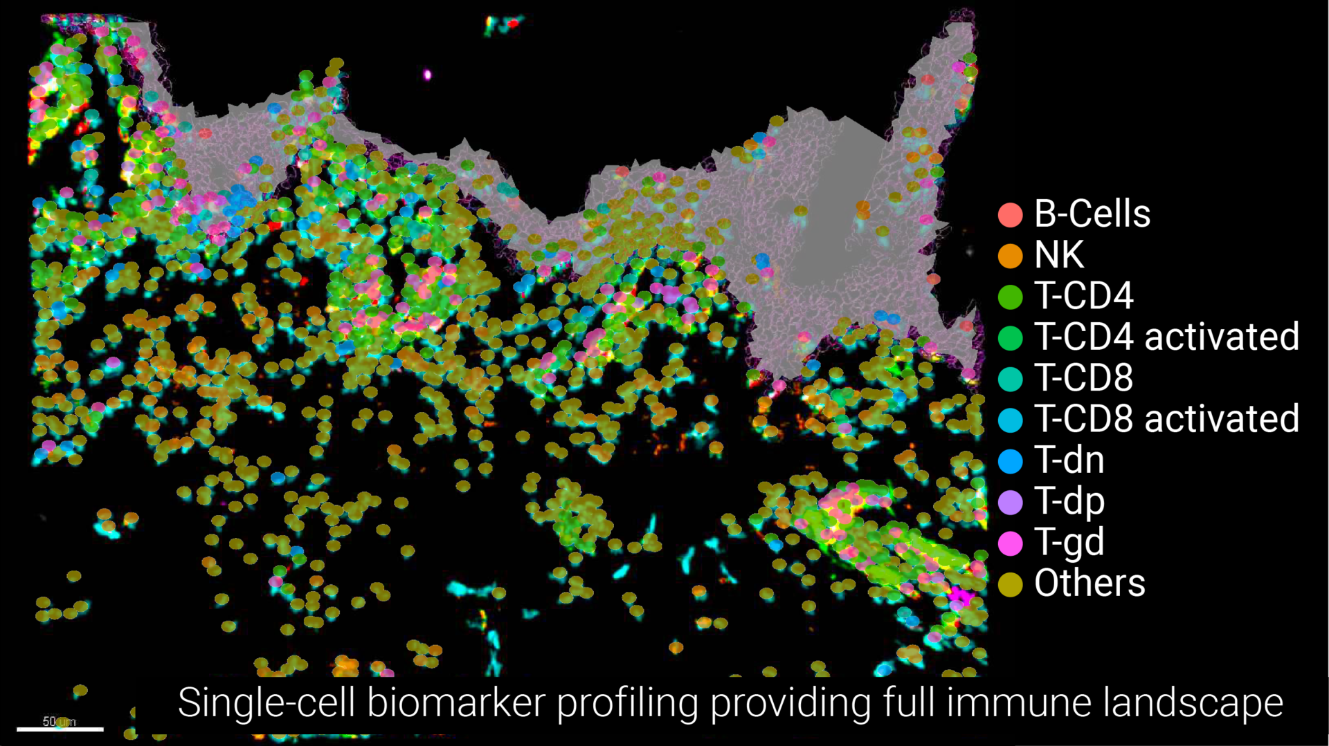
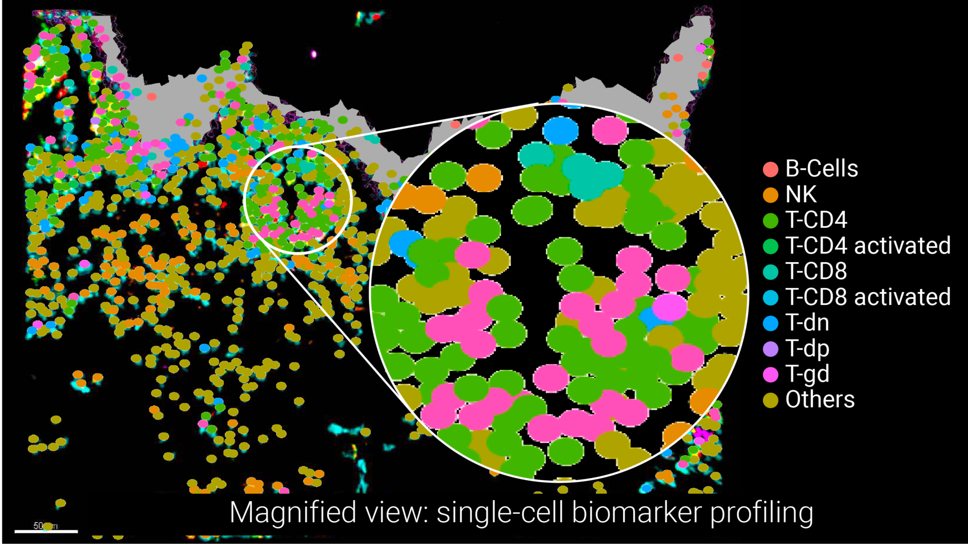
Powered by multiplexed 3D imaging, MANTIS® automatically projects a representative digital immune landscape, while enabling interactive gating of anatomical regions and concomitant single-cell data quantification of biomarkers. This unique service provides researchers with 3D multiplexed imaging of human skin tissue and enables the unbiased complete immune profiling of skin tissue within its spatial context.
Unbiased automated analysis
Gain new insights into human immune response
Genoskin’s 3D multiplexed image acquisition is associated with in-house automated image processing. Our interactive and user-friendly analytical software allows for unbiased analysis. The proprietary interface generates a digitalized version of the skin immune landscape, which enables single-cell quantitative data visualization at unprecedented levels. The MANTIS® platform provides groundbreaking insights into the human skin’s immune response to vaccines, drugs and cosmetics.
Full immune profiling with 3 pre-designed panels

The MANTIS® spatial biology platform integrates seamlessly into our existing data generation platforms such as the Injection Site Reactions (ISR) Platform for vaccine and drug development studies, as well as into our range of real human skin models that provide live human skin response, such as HypoSkin®, InflammaSkin® or NativeSkin®. Dive deeper into tissue characterization upon drug administration thanks to our three pre-designed panels.
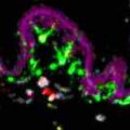
Myeloid cells
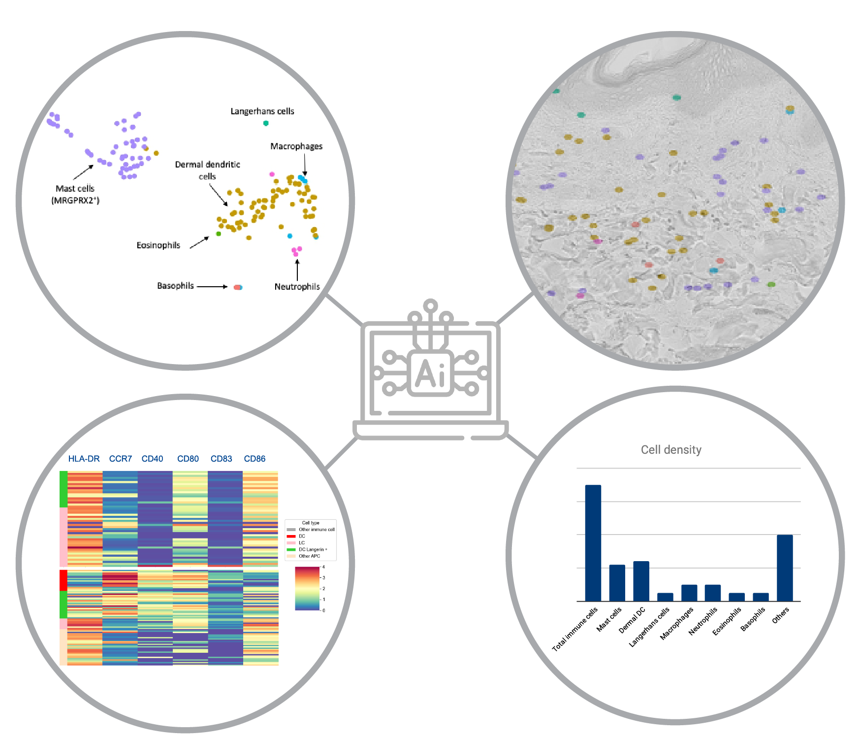
Analyze Langerhans cells, dendritic cells, macrophages, mast cells, eosinophils, basophils, neutrophils.
CD45 – CD1c – CD207 – Chymase – pan-HLA – Siglec 8 – CD123 – MPO – CD68
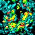
Lymphoid cells
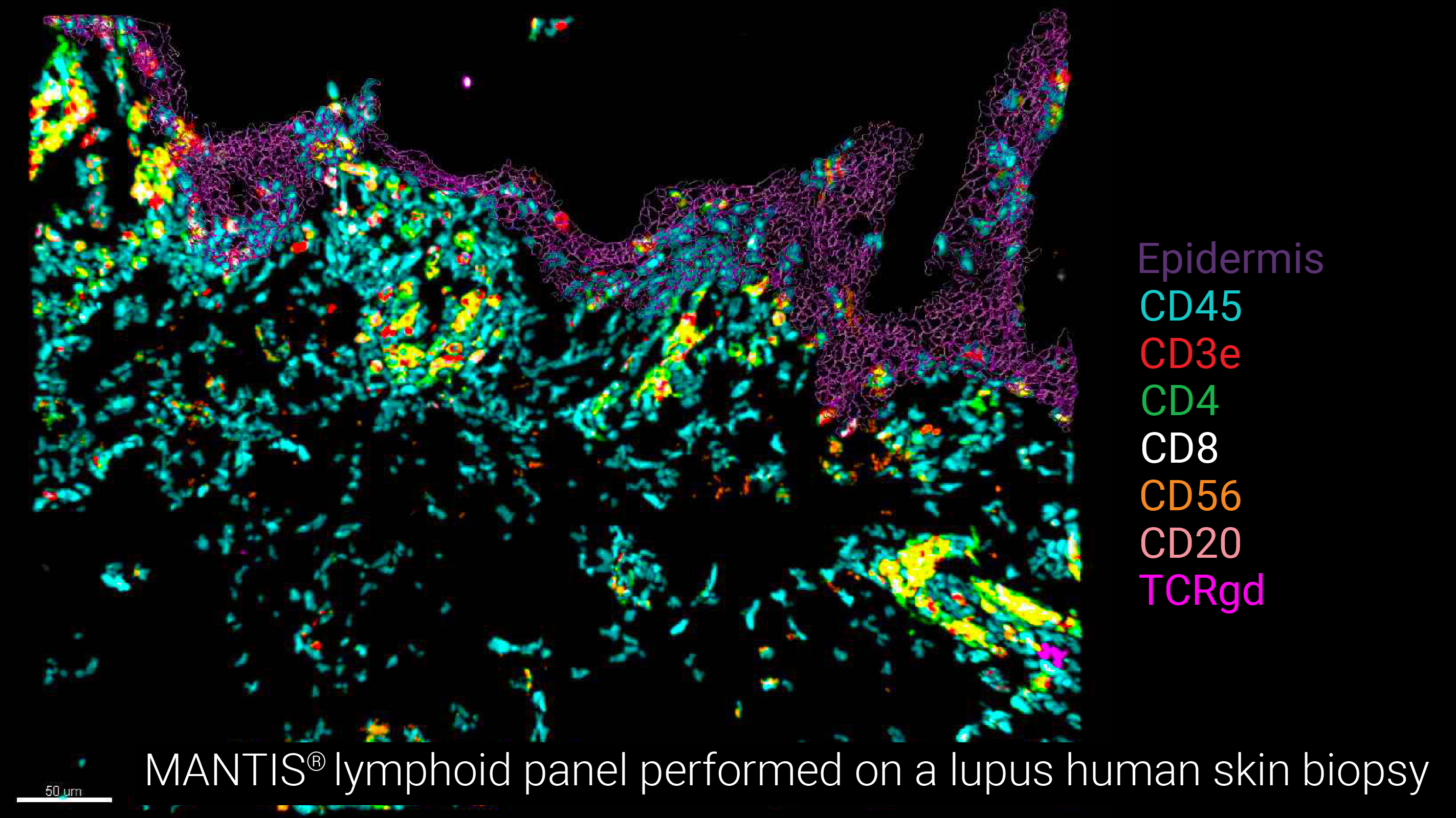
Analyze T helper cells, Cytotoxic T cells, B cells, NK cells, gd T cells.
CD45 – CD3e – CD4 – CD8a – CD57 – CD20 – CD31 – TCRgd
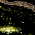
Antigen-presenting cell (APC) activation
Analyze Langerhans cells, dendritic cells, macrophages and their activation status.
CD45 – CD1c – CD207 – pan-HLA – CD80 – CD83 – CD86 – CD40 – CCR7
A flexible & interactive analytical spatial skin biology platform

The MANTIS® spatial skin biology platform acquires the data of 12+ markers in one single acquisition using a classical confocal microscope coupled with a powerful user-friendly analytical interface. The method also leverages the natural autofluorescence of structural elements to model the architecture of the skin and easily position anatomical sections.


