Skin Models
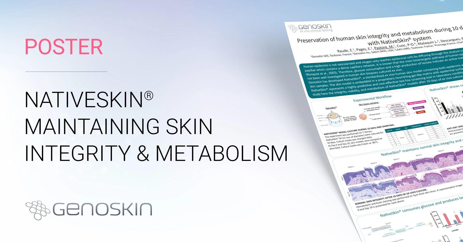
Preservation of human skin integrity and metabolism during 10 days in culture with NativeSkin® system
Research by E. Raude1, E. Pagès1, M. Pastore1, P.-O. Cuoc2, L. Malaquin3, P. Descargues1
1Genoskin, 2Promega, 3LAAS-CNRS
Scientific Context
Human epidermis is not vascularized and oxygen only reaches epidermal cells by diffusing through the stratum corneum or from the dermal papillae which contains a dense capillary network. It is known that the main bioenergetic pathway of human epidermis is anaerobic glycolysis (Ronquist et al., 2003). Therefore, glucose consumption and high production of lactate indicate an active metabolism in the skin. This pathway has not been well investigated in human skin biopsies cultured ex vivo. Genoskin has developed NativeSkin®, a standardized ex vivo human skin model containing both epidermis and dermis, produced with discarded skin samples. The skin model is embedded in a proprietary nourishing gel-like matrix with the epidermal surface left in direct contact with the air. NativeSkin® represents a highly predictive and cost-effective last-line screening tool in laboratory conditions prior to in vivo clinical evaluations. We study here the integrity, viability, and metabolism of NativeSkin® models after ten days of ex vivo culture.
NativeSkin® shows cell proliferation and no apoptosis at day 10
Anti-Ki67 and anti-active Caspase-3 immunostainings were performed on 5µm thickness skin slices to assess cell proliferation and apoptosis. Representative images were presented. Quantifications were performed on 6 representative pictures of the epidermis.
37% of cell proliferation were maintained after 10 days of ex vivo culture. Depending on the donor, levels of proliferation were from 22 to 69% of levels just after surgery. Levels of apoptosis marker were low after 10 days of ex vivo culture, with only 0.15 to 2% of the total epidermis area stained.

Anti-KI67 and anti-active caspase-3 immunostainings and quantification.
Please fill in the form below to access our poster.
After submitting this form, you will receive an email with the link to access our poster.
Comments are closed.


