
HairSkin® ex vivo skin model
Study hair and scalp biology in a relevant human tissue context
HairSkin®: ex vivo human skin model with hair follicles
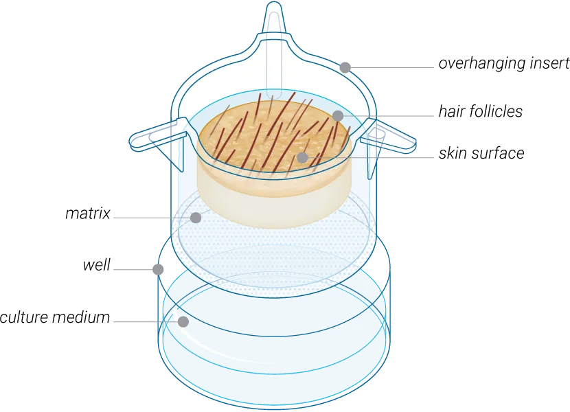
HairSkin® is an ex vivo human skin model with hair follicles and mature pilosebaceous units, designed for pharmaceutical, cosmetic, dermatology, and haircare research. By preserving natural architecture of human skin, hair follicles, sebaceous glands, and immune cells, HairSkin® helps generate relevant human proof-of-concept data to evaluate hair follicle biology, sebaceous gland function, and the local efficacy and toxicity of topical treatments, drugs, and cosmetics.
The Challenge
Hair care and hair drug development require models that can capture the complexity of human hair and scalp biology. Yet many existing models fail to reproduce the biology of the hair follicle, making it difficult to generate data that truly reflects human response, especially when studying scalp health, inflammation, and follicle-related disorders.
HairSkin® can help overcome these challenges by providing an ex vivo human skin model with mature hair follicles and pilosebaceous units. By preserving the natural architecture of human skin, scalp tissue, and its appendages, HairSkin® enables researchers to evaluate products, ingredients, and therapeutic candidates in a more relevant human biological context.
For cosmetic, personal care, and pharmaceutical companies, HairSkin® supports more confident decision-making by providing actionable human data earlier in the development process, from scalp health preservation to anti-inflammatory strategies and hair-focused innovation.
Applications
Hair follicle biology research
Drug efficacy & toxicity assessment
Skin condition models
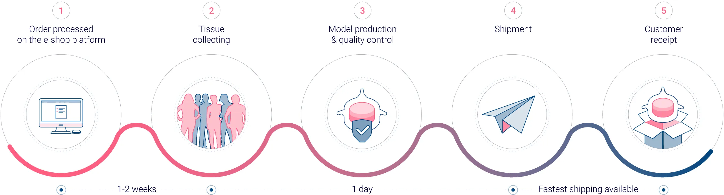
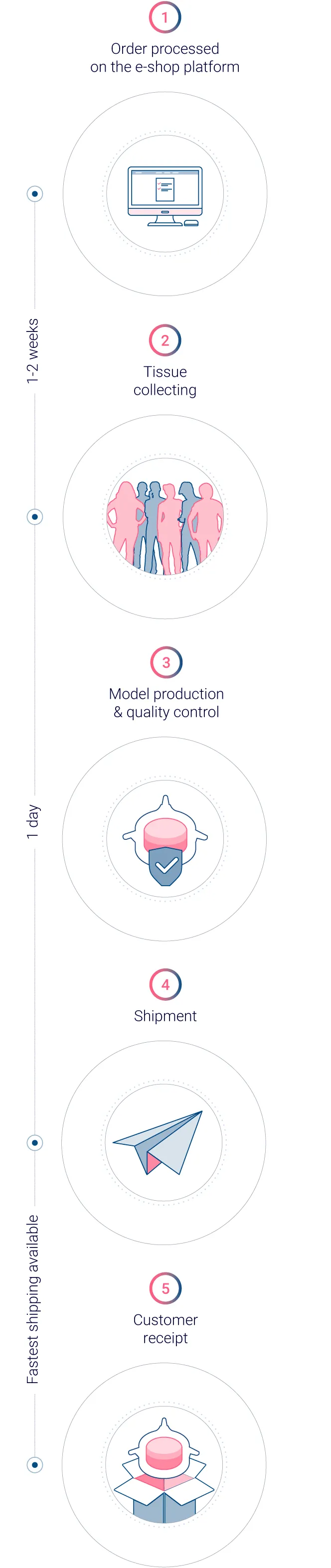
Standardized production process for actionable data
Genoskin implements a standardized workflow to produce reliable, consistent, and ready-to-use human skin models. Thanks to a patented technology, we ensure that every model is standardized, providing accurate and reproducible results.
Biological validation data
Viability of the hair follicle
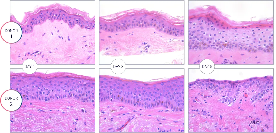
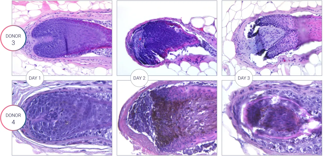
Figure: H&E staining showing the hair follicle characterization during the culture. Viability of the hair follicle measured over 3 days for different donors.
Hair follicle characterization
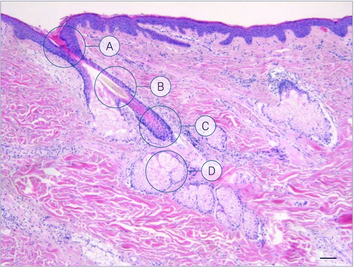
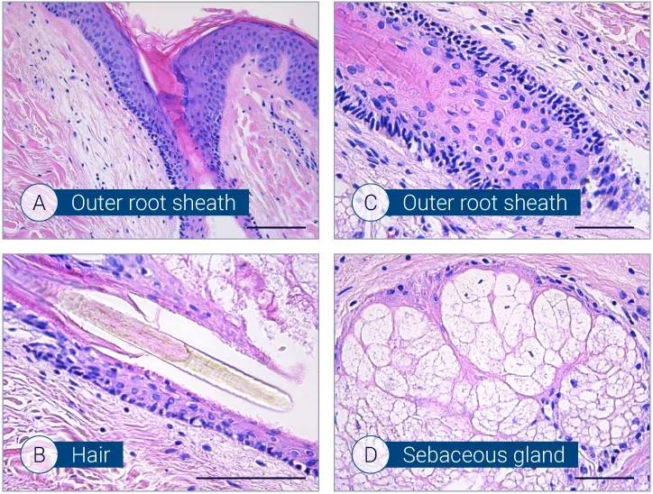
Figure: H&E staining showing characterization of hair follicles on HairSkin® models. 5 µm-thick FFPE sections stained with H&E. Scale bar: 100µm.
The evolution of the wound without treatment has been followed over 11 days by histology and immunofluorescence. The data are showing the closing of the wound over time.
Characterization of immune cells and nerve endings
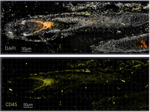
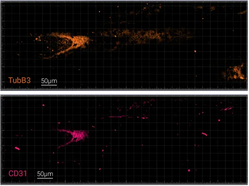
Figure: Structural and activation markers analysis performed on a tissue cryosection with a thickness of 40µm: DAPI (nucleus), CD45 (immune cells), Tubulin β3 (nerve endings), CD31 (blood vessels).

Order HairSkin® access
Try it in your own lab
Order our shippable HairSkin® models via our online platform and get them delivered straight to your lab ready-for-use, along with a step-by-step user manual.