
Biostabilized Hs Skin | Preclinical Hs Research
Hidradenitis suppurativa research studies in ex vivo human HS skin
Preclinical hidradenitis suppurativa studies using a New Approach Methodology

Human-relevant assays to evaluate therapeutic responses in hidradenitis suppurativa (HS) tissue
Move beyond simplified disease models with NAMs. Investigate therapeutic response directly in human hidradenitis suppurativa tissue.
Genoskin is a Contract Research Organization (CRO) which provides access to human HS skin collected from affected donors and transformed into functional, biostabilized ex vivo models for controlled therapeutic testing. Study how your drug candidate affects the complex biology of diseased human skin—from inflammatory signaling and cytokine release to tissue morphology and molecular responses.
What is hidradenitis suppurativa, and why study it ex vivo?
Hidradenitis suppurativa (HS), also known as acne inversa, is a chronic inflammatory skin disease of the hair follicle that causes painful nodules, abscesses, draining tunnels and scarring. It affects an estimated 1–4% of the population in Western countries, yet its immunobiology remains poorly understood and treatment options are limited. Because HS is a solely human disease that no animal model fully reproduces, patient-derived HS skin offers one of the most direct ways to evaluate new therapies before clinical trials.
From patient-derived HS tissue to therapeutic response
HS is driven by complex interactions between the epidermis, hair follicles, immune cells, inflammatory mediators and the surrounding tissue microenvironment. Reproducing this biology in conventional in vitro systems remains challenging.
With Genoskin, you can investigate your therapeutic in human HS tissue.
Real human HS disease tissue
Work with skin tissue, including lesional tissue, from patients with HS to study your therapeutic in the context of the disease it is designed to treat.

Functional ex vivo model
Biostabilized HS skin preserves native tissue architecture and relevant cellular components, so you can move from static tissue characterization to controlled therapeutic exposure.

Multiple complementary readouts
Connect cytokine, histological, immune, molecular and spatial data to understand how treatment modifies diseased human skin.

Customized study design
Build the experiment around your therapeutic, target and research question.
Test your therapeutic directly in human HS skin
What is it and how does it work?
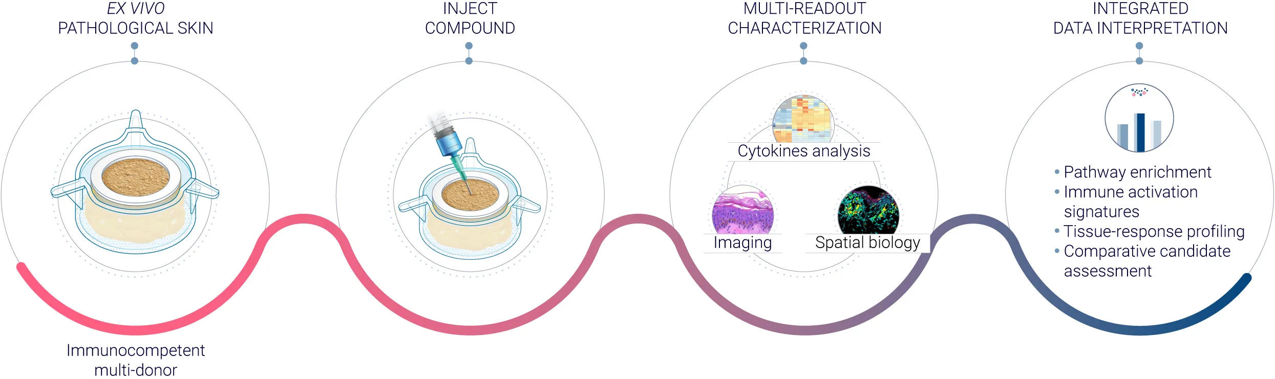
Native HS skin tissue is maintained in our ex vivo biostabilization platform, allowing controlled dosing (e.g., subcutaneous injection or topical application) and multi-day treatment response tracking. You can evaluate your small molecules, biologics or topical candidates on diseased human HS skin tissue.
Key benefits
Test efficacy in relevant human disease tissue before clinical trials
Observe treatment-associated changes over an optimal 3- to 7-day readout window
Benchmark your candidates against established reference therapies such as Humira® (adalimumab)
Treatment-response studies
Compare treated and untreated human HS tissue and evaluate treatment-associated changes across relevant biological endpoints.
Potential applications include:
- Therapeutic efficacy assessment
- Dose-response evaluation
- Candidate or formulation comparison
- Reference-treatment benchmarking
- Mechanism-of-action investigation
- Biomarker identification and characterization
- Translational studies supporting therapeutic development
Want more research like this in your inbox? Subscribe to our newsletter →
A functional response to an established HS therapy
Treatment-associated changes observed following Humira® (adalimumab) exposure
To evaluate whether biostabilized human HS skin could capture a pharmacological response, HS models were treated with Humira® as a reference therapy. Treatment produced measurable changes in inflammatory cytokines and tissue morphology, demonstrating the utility of biostabilized HS skin for investigating therapeutic response in human disease tissue. Complementary macroscopic and spatial analyses characterize tissue preservation and the immune landscape of the model.
Measurable suppression of inflammatory signaling in diseased human skin
Treatment with an established HS therapy produced measurable changes in inflammatory cytokines and tissue morphology, demonstrating the utility of biostabilized HS skin for investigating therapeutic response in human disease tissue.

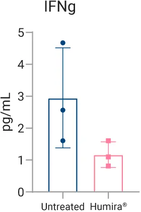
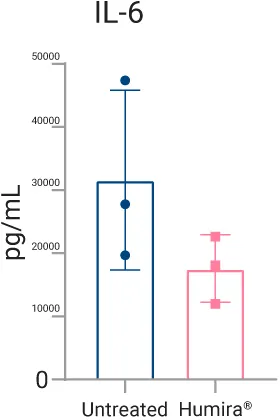
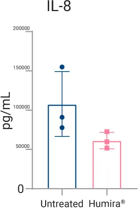
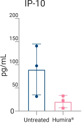
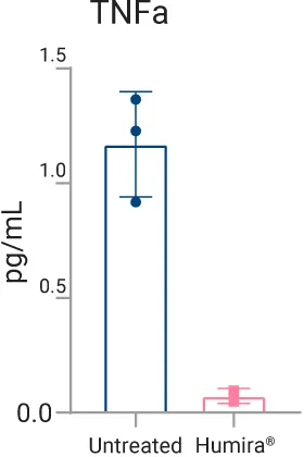

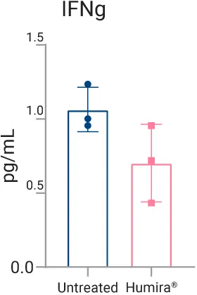
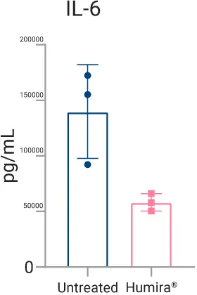
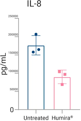
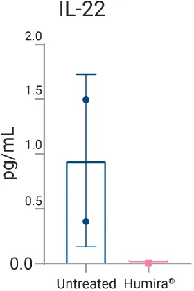
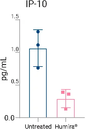
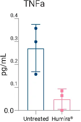
Quantification of pro-inflammatory cytokine concentrations (pg/mL) measured via Meso Scale Discovery (MSD) multiplex immunoassay in cell culture supernatants collected from biostabilized human HS skin models on Day 3 and Day 7.
(A) Day 3 supernatant levels of IFN-γ, IL-6, IL-8, IP-10, and TNF-α comparing untreated HS skin (blue) against Humira®-treated skin (pink).
(B) Day 7 supernatant levels showing sustained suppression of IFN-γ, IL-6, IL-8, IL-22, IP-10, and TNF-α following Humira® treatment. Bars represent group mean values, with error bars indicating standard deviation (SD) across individual experimental replicates.
Humira® reduces inflammation-driven damage in HS models
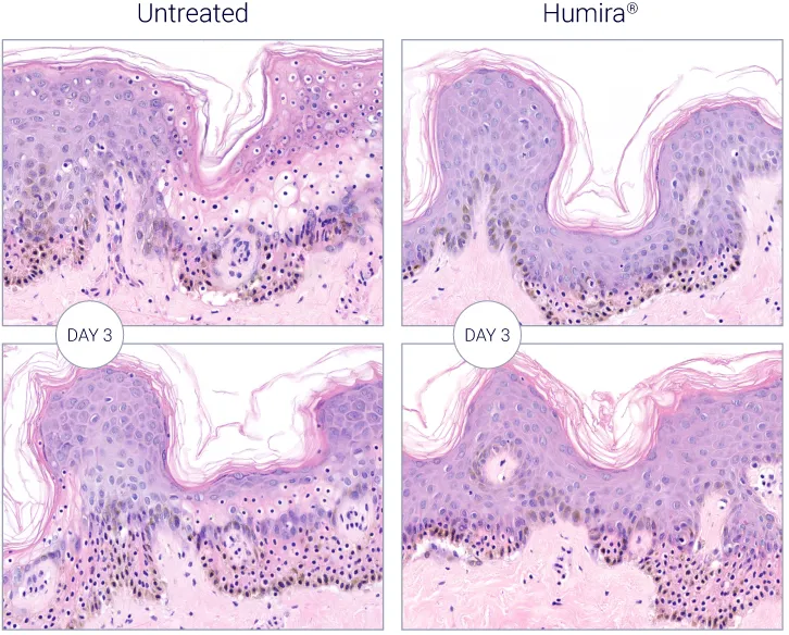
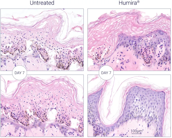
Longitudinal assessment of epidermal integrity and tissue architecture by H&E staining. Compared with untreated controls, Humira® (adalimumab) reduced inflammation-driven tissue damage and preserved overall epidermal architecture at Days 3 and 7. Reduced pyknotic cell formation further indicates improved tissue preservation following treatment. Representative images from two replicates per condition are shown.
Ex vivo macroscopic characterization and biostabilization of human HS skin models

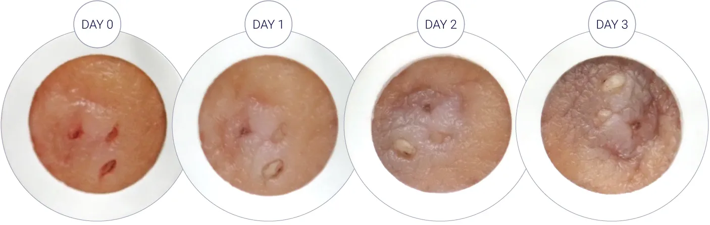

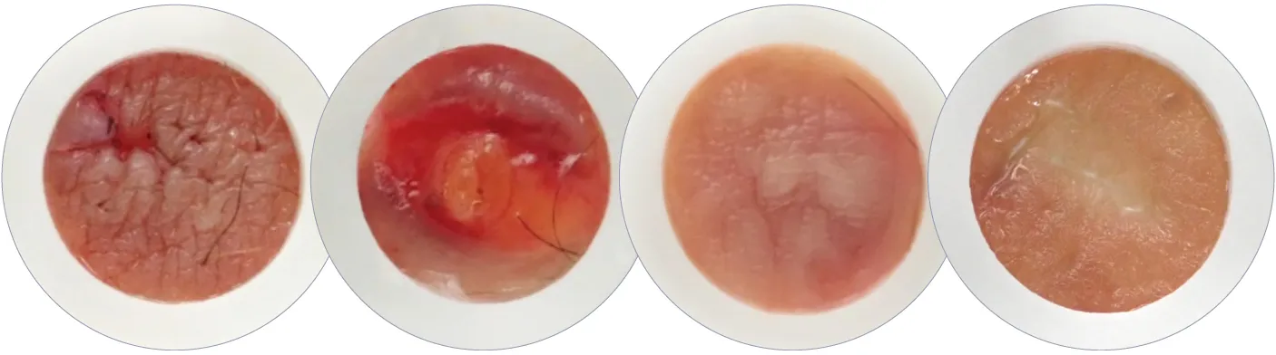
A – Daily macroscopic imaging (Day 0 through Day 7) evaluating surface integrity and tissue preservation of biostabilized ex vivo HS skin models under untreated control conditions.
B – Examples of lesional tissue from multiple donors.
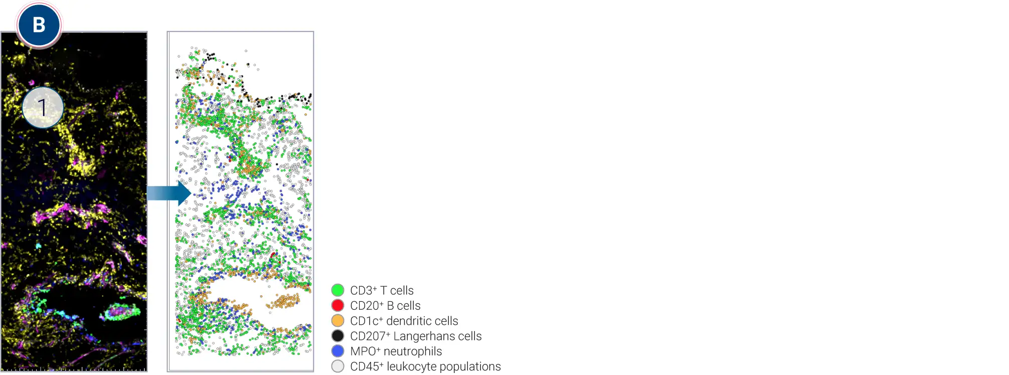
Spatial biology and digital cell mapping of immune infiltrates in ex vivo HS lesions
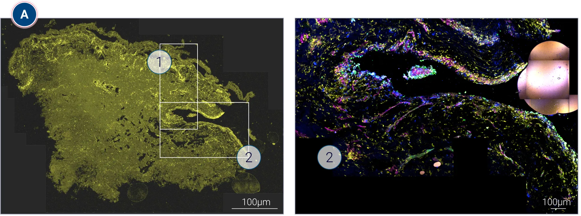
(A) High-multiplex digital imaging mapping specific markers—including CD3+ (T cells), CD20+ (B cells), CD1c+ (dendritic cells), CD207+ (Langerhans cells), MPO+ (neutrophils), and CD45+ immune populations to evaluate immune cell populations and location in the disease tissue.
(B) Corresponding digital single-cell spatial map illustrating spatial localization of phenotyped leukocyte subsets: CD3⁺ T cells (green), CD20⁺ B cells (red), CD1c⁺ dendritic cells (orange), CD207⁺ Langerhans cells (black), MPO⁺ neutrophils (blue), and other CD45⁺ leukocyte populations (white). Scale bar = 400 µm
Learn more about our spatial biology platform.
Need the tissue instead? Work with human HS samples in your own laboratory.
For researchers who want to run their own experiments, hidradenitis suppurativa tissue samples are available in four research-ready formats: live biostabilized skin models, frozen biopsies, FFPE blocks and tissue in RNAlater™.
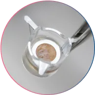
11 mm biostabilized HS models
Live human HS tissue prepared as 11 mm biostabilized skin models for ex vivo experimentation in your laboratory.
Ideal for: functional studies, treatment experiments and tissue-based assays requiring viable tissue and intact immune cells to capture biologically relevant treatment responses.
Availability: Europe only

4 mm frozen HS biopsies
Research-ready 4 mm frozen punch biopsies for downstream molecular and biochemical analysis.
Ideal for: molecular profiling, biomarker discovery, gene expression, proteomic analysis, histology, immunohistochemistry, pathway characterization and other downstream tissue-based research applications.
Availability: Worldwide
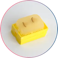
FFPE HS tissue blocks
Formalin-fixed, paraffin-embedded human HS tissue for morphology and tissue biomarker studies.
Ideal for: histology, IHC, immunofluorescence and target-expression analysis.
Availability: Worldwide
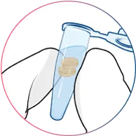
HS tissue in RNAlater™
Human HS samples preserved in RNAlater™ for downstream RNA and transcriptomic analysis.
Ideal for: gene-expression studies, bulk RNA sequencing and transcriptomic biomarker discovery.
Availability: Worldwide
Frequently asked questions
Can I test my compound on the HS skin model?
Can Genoskin perform the testing for me?
Yes. In addition to supplying HS tissue products, Genoskin is a CRO that offers customized research services using biostabilized human HS models. Study designs can be tailored to your research objectives, including treatment response, mechanism-of-action and biomarker studies.
What is the difference between the HS skin model and HS tissue samples?
The biostabilized HS skin model is designed for functional and treatment experiments using viable human HS tissue. It can be used to evaluate tissue responses following treatment and investigate mechanisms of action, targets and biomarkers.
HS tissue samples (frozen biopsies, FFPE tissue blocks) are provided in formats optimized for downstream molecular, biochemical or histological analyses, depending on the product.
What can HS tissue be used for? What types of analyses can be performed using HS tissue?
HS tissue supports research into HS disease biology, inflammatory mechanisms, biomarkers, molecular pathways and therapeutic responses. Depending on the tissue format, applications include molecular profiling, gene expression analysis, histology, immunohistochemistry, immunofluorescence, biomarker analysis and functional treatment studies.
Depending on the tissue format, HS tissue can be used for:
- Gene expression and transcriptomic analysis
- Molecular profiling
- Protein and biomarker analysis
- Histology
- Immunohistochemistry (IHC)
- Immunofluorescence (IF)
- Target and pathway characterization
- Tissue morphology assessment
What HS tissue products does Genoskin offer?
What donor and disease information is provided with HS tissue samples?
Genoskin can provide available donor information collected with the tissue sample, including reported medical conditions, treatments, allergies, and HS disease severity (HS stage). The information provided depends on the availability of the relevant donor data and can help support sample selection and interpretation of research results.
How much HS tissue is available?
The quantity of tissue available varies depending on the size and characteristics of the surgical specimen received. For HS skin models, an average of at least 10 models can be generated from each surgical specimen.
Does HS tissue vary between donors?
Yes. Human HS tissue naturally varies in morphology, disease characteristics (including Hurley stage) and biological features between donors. This variability should be considered when designing experiments and interpreting results.
Can I purchase HS tissue products directly online?
Selected HS tissue products are available through the Genoskin eShop. Product availability may vary based on current tissue supply. For specific donor requirements, larger quantities or customized studies, please contact Genoskin. The 11 mm models are only available for sale in European countries. The FFPE tissue blocks, frozen biopsies and HS tissue in RNAlater™ are available globally.
Want more research like this in your inbox? Subscribe to our newsletter →