
InflammaSkin® | Preclinical psoriasis research
Human Ex Vivo Psoriasis Model for Preclinical Drug Efficacy Studies
Why use a human psoriasis model instead of standard preclinical models?
Today’s preclinical skin models can mimic the inflammatory features of psoriasis, but they often lack tissue complexity and immune cell diversity, and most cannot receive a subcutaneous injection. Developed by Genoskin, a CRO specializing in New Approach Methodologies (NAMs), InflammaSkin® closes that gap with live, immunocompetent human skin, for both drug development and psoriasis biology research.
| Criteria | InflammaSkin® human psoriasis model | Typical preclinical skin models |
|---|---|---|
| Tissue | ✓Real human epidermis and dermis, plus adipose tissue when needed | ~Limited tissue complexity |
| Immune cells | ✓The structural and immune cell types of native skin | ~Limited immune cell diversity |
| Inflammation | ✓Resident T cells activated in situ, Th17/Th1-driven | ~Mimics inflammatory features |
| Subcutaneous dosing | ✓Subcutaneous and intradermal injection | ✗Generally not possible |
| Modalities | ✓Biologics and small molecules | — |
InflammaSkin®: A T cell-driven human skin psoriasis model
InflammaSkin® is the only T cell-driven psoriasis model built on real ex vivo human skin. Test biologics and small molecules applied topically, systemically or by subcutaneous injection, and get immune-relevant human data before clinical trials.
- Real human epidermis and dermis, with optional adipose tissue for injections
- Resident T cells activated in situ to a Th17/Th1 psoriasis phenotype
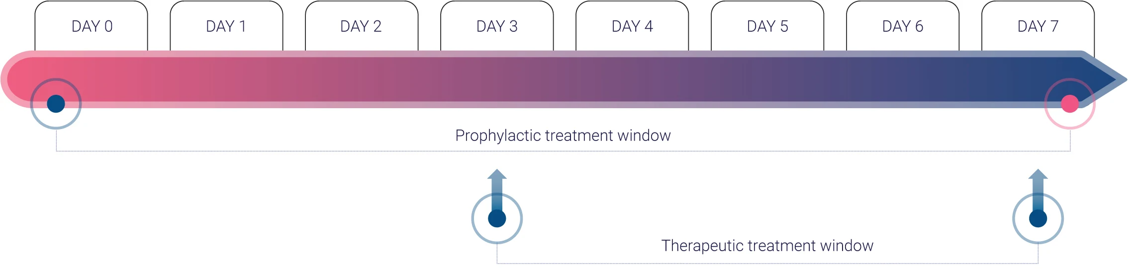
- Prophylactic (Day 0) or therapeutic (Day 3) dosing over a 7-day window
T cell driven
Currently the only ex vivo psoriasis skin model on the market with T cell-driven Th17/Th1 inflammation.

Immunocompetent
Alive and functional, with the structural and immune cell types and appendages of native human skin.

Reproducible
Standardized human skin explant models for consistent, immune-relevant inflammation readouts.

Versatile
Suited to prophylactic and therapeutic biologics and small molecule drugs.
Psoriasis, a condition with no cure
Helping science move forward with NAMs
Psoriasis is an immune-mediated skin disease that affects over 125 million people worldwide according to the World Psoriasis Day consortium. The condition has a big impact on patients who are also at risk of experiencing a range of associated diseases, such as metabolic syndrome, stroke, heart attack, cardiovascular disease and depression. About 40% of patients with psoriasis develop psoriatic arthritis. To date, anti-psoriasis drugs don’t provide a cure, even though biologics have helped improve symptoms. Genoskin is a CRO that aims to provide drug developers with innovative solutions to help them better serve patients.
Design your preclinical psoriasis study
Evaluate therapies targeting key psoriasis pathways, including T cells and cytokines such as IL-17, IL-22, and TNF-α, in early discovery or confirmatory studies.
The InflammaSkin® model empowers researchers to assess the efficacy of anti-inflammatory treatments with precision. This model is meticulously designed to evaluate therapies targeting key psoriasis pathways, including T cells and cytokines such as IL-17, IL-22, and TNF-α.
Whether you’re focusing on early intervention with prophylactic compounds (Day 0 administration) or addressing established inflammation with therapeutic treatments (Day 3 administration), InflammaSkin® adapts to your research needs.
Administration routes
Readouts included
Results analyzed by our experts in human skin and immunology.
Validation data: An experimental psoriasis model with immune-relevant readouts
Multi-parametric readouts reveal cytokine profiles, therapeutic response and immune cell shifts
Subcutaneous administration of anti-TNF-α drives dose-dependent cytokine suppression in InflammaSkin®

Cytokine changes following anti-inflammatory treatment can be measured in InflammaSkin®

A.B. All cytokines for the HypoSkin® control are down-regulated in comparison to the Hypo-InflammaSkin control → there is significant inflammation induction in the Hypo-InflammaSkin models at day 7. Additionally, we observed a 300-fold increase in IL-17 levels in Hypo-InflammaSkin compared with HypoSkin®, confirming effective induction of the inflammatory response.
Betamethasone treatment reduced the secretion of several cytokines compared with untreated Hypo-InflammaSkin controls, including IFN-γ, IL-10, IL-17A, IL-22, IL-8, MIP-3α, and TNF-α.
Characterization of histological changes after induction of Th17/Th1 inflammation
Hematoxylin and eosin staining was performed on 5 µm thick skin cross-sections. The histological analysis of the InflammaSkin® model shows epidermal anomalies that are characteristic of psoriasis, in contrast to non-inflamed ex vivo skin models and the same model treated with the pro-inflammatory cocktail. All images are representative of the whole sample. Scale bar is 100 µm.
No induction
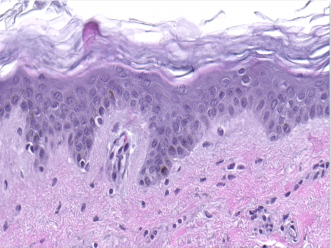
Healthy ex vivo human skin model cultured under basal conditions.
No T cell activation
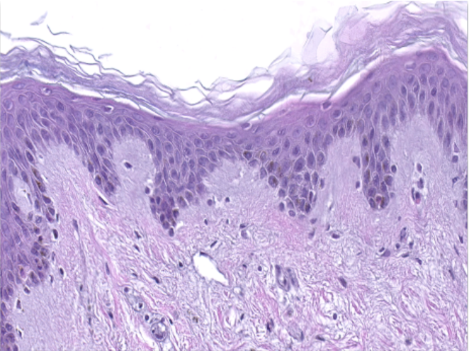
Skin model cultured in the presence of a Th17/Th1 polarization cocktail but without T cell activation.
InflammaSkin®
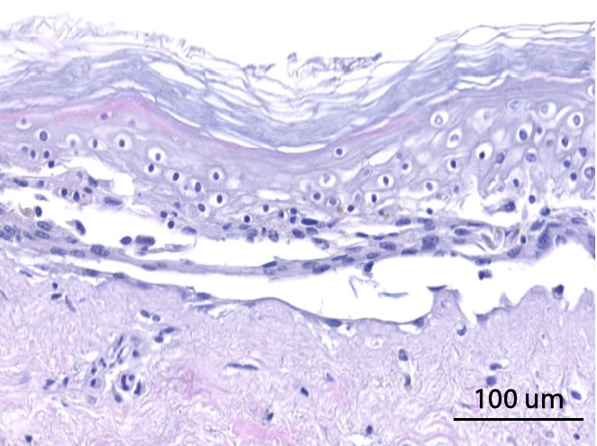
InflammaSkin® Model with T cell activation cocktail, cultured in the presence of a Th17/Th1 polarization cocktail for 7 days.
Expression of pro-inflammatory markers in InflammaSkin®
Epidermal activation is demonstrated by the over-expression of the psoriasis-associated pro-inflammatory markers Keratin 16 (K16) and Psoriasin (S100A7). Anti-S100A7 and anti-K16 immunofluorescence staining was performed on 5 µm thick skin cross-sections. Red and cyan signals indicate a positive signal and DAPI-counterstain nuclei, respectively. The dotted white lines represent the epidermal-dermal junction. Epidermal and dermal compartments are located above and below the dotted line respectively. All images are representative of the whole sample. Scale bar is 100 µm.
No induction
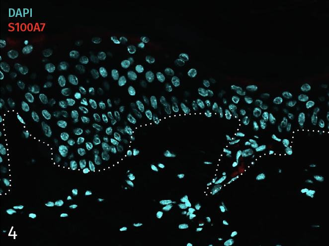
Presence of pro-inflammatory marker Psoriasin (S100A7) in a healthy ex vivo skin model cultured under basal conditions.
No T cell activation
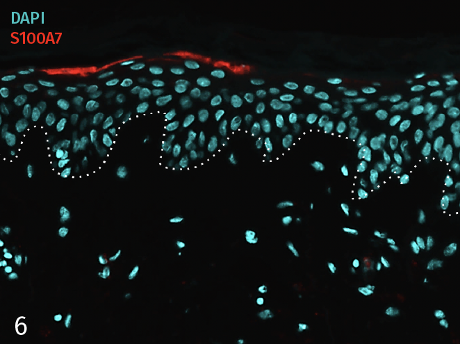
Presence of pro-inflammatory marker Psoriasin (S100A7) in a skin model cultured in a Th17/Th1 polarization cocktail but without T cell activation.
InflammaSkin®
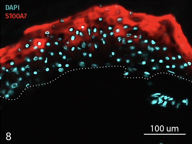
Presence of Psoriasin (S100A7) in the InflammaSkin model with T cell activation, cultured in a Th17/Th1 polarization cocktail for 7 days.
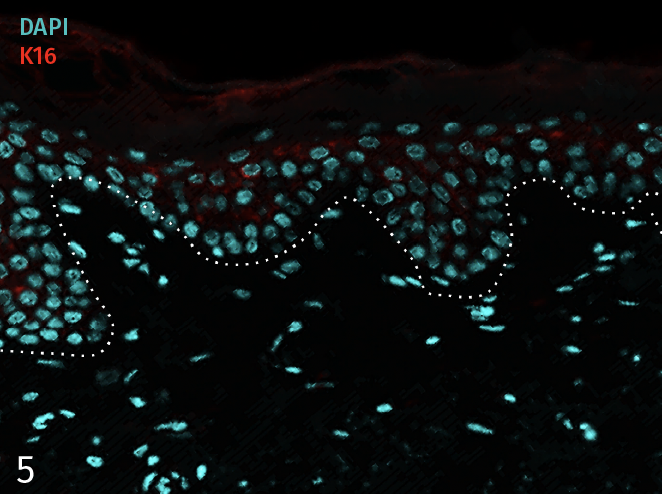
Presence of pro-inflammatory marker Keratin 16 (K16) in a healthy ex vivo skin model cultured under basal conditions.
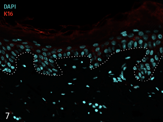
Presence of pro-inflammatory marker Keratin 16 (K16) in a skin model cultured in a Th17/Th1 polarization cocktail, without T cell activation.
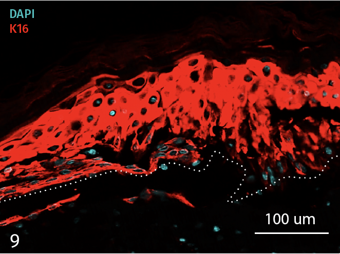
Presence of Keratin 16 (K16) in the InflammaSkin model with T cell activation, cultured in a Th17/Th1 polarization cocktail for 7 days.
CD69 and Ki-67 expression highlights enhanced T cell activation and proliferation in InflammaSkin®

Robust quality control at every stage of the study
From donor selection to analysis: A controlled workflow
The results obtained from our InflammaSkin® platform are analyzed by a team of experts in human skin and immunology, who work with cutting-edge tools to help you obtain the results to help your research move forward. Our experts master the traditional, standard tests as well as the latest technology to assess skin inflammation, epidermal activation and tissue integrity. We focus on generating unbiased and accurate human data to support safer and faster drug development, backed by our ethical sourcing and quality assurance standards. Below you’ll find an illustration of our psoriasis research study process.


Download our InflammaSkin® flyer
Discover InflammaSkin®, a human ex vivo psoriasis model for preclinical efficacy testing of topical and injectable drug candidates. It reproduces key features of psoriasis-like inflammation through T cell activation and Th17/Th1 polarization, while preserving the tissue complexity and immune cell diversity of human skin.
Flyer developed by Genoskin’s in-house scientific team
Immediate access upon form submission
Your information is confidential and protected
Get the InflammaSkin® flyer
Frequently asked questions
What is InflammaSkin® and how is it used in psoriasis research?
What types of psoriasis treatments can be tested with InflammaSkin®?
What psoriasis biomarkers and readouts can be measured?
Can InflammaSkin® be used to assess treatment efficacy?
Can InflammaSkin® models be purchased for use in my own laboratory?
What makes InflammaSkin® different from reconstructed psoriasis skin models?
Does InflammaSkin® contain functional human T cells?
Can InflammaSkin® be used for dose-response studies?
Why use a human ex vivo psoriasis model during preclinical development?
Scientific publications using our technology & services
See how others use our Inflammaskin® model and services
July 2024 - Press Release - Scinai Immunotherapeutics announces promising results in an in-vivo proof-of-concept psoriatic human skin model
PR – July 15th, 2024.
The results confirm and build upon previous reported results from ex-vivo studies done with human skin specimens (conducted by Genoskin) and in a plaque psoriasis in vitro model with human skin tissue grown in a dish.
June 2024 - Challenges in psoriasis research: a systematic review of preclinical models.
Published in Karger – June 10, 2024. https://doi.org/10.1159/000538993.
Ana Ubago-Rodríguez, María I. Quiñones-Vico, Manuel Sánchez-Díaz, Raquel Sanabria-de la Torre, Álvaro Sierra-Sánchez, Trinidad Montero-Vílchez, Ana Fernández-González, Salvador Arias-Santiago.
December 2023 - The development of human ex vivo models of inflammatory skin conditions.
Published in National Library of Medicine – December 8, 2023. Int J Mol Sci. 2023 Dec;24(24):17255. doi:10.3390/ijms242417255.
Eddy Hsi Chun Wang, Rebecca Barresi-Thornton, Li-Chi Chen, Maryanne Makredes Senna, I-Chien Liao, Ying Chen, Qian Zheng, Charbel Bouez
December 2023 - Press Release - Scinai announces promising results in a psoriatic human skin model
PR – Dec 12th, 2023
The study, conducted by Genoskin, a pioneering French biotechnology company, aimed to evaluate the anti-inflammatory effects of Scinai’s NanoAbs. Genoskin’s proprietary human skin models were induced for expression of plaque psoriasis symptoms to enable ex‑vivo examination of the therapeutic effects of drugs targeting underlying mechanisms in the pathogenesis of plaque psoriasis, particularly the IL-17 family of pro-inflammatory cytokines. This disease-induced skin model reproduces key features of plaque psoriasis tissue morphology as well as the cytokine profile associated with the inflammatory state of plaque psoriasis lesions. Genoskin’s model has been successfully validated as a reliable ex-vivo system for testing drugs aimed at plaque psoriasis.
February 2021 - Calcipotriol/Betamethasone Dipropionate foam inhibits Th17 cytokine secretion and improves epidermal barrier markers in a human Th17 skin inflammation model
- Calcipotriol/Betamethasone Dipropionate Foam Inhibits Th17 Cytokine Secretion and Improves Epidermal Barrier Markers in a Human Th17 Skin Inflammation Model
- Published in Dermatologic Therapy – 2021 Feb;11(1):265-274
- Paola Lovato, Li Jiang, Josephine Hebsgaard, David A Ewald, Hanne Norsgaard
- Published in Dermatologic Therapy – 2021 Feb;11(1):265-274
October 2020 - Development and characterization of a human Th17 driven ex vivo skin inflammation model
- Development and characterization of a human Th17‐driven ex vivo skin inflammation model
- Published in Experimental Dermatology – Oct. 2020;29(10):993-1003.
- Claire Jardet, Anthony David, Emilie Braun, Pascal Descargues, Jean-Louis Grolleau, Josephine Hebsgaard, Hanne Norsgaard, Paola Lovato
- Published in Experimental Dermatology – Oct. 2020;29(10):993-1003.
January 2020 - Translational drug discovery and development with the use of tissue-relevant biomarkers: Towards more physiological relevance and better prediction of clinical efficacy
- Translational drug discovery and development with the use of tissue-relevant biomarkers: Towards more physiological relevance and better prediction of clinical efficacy
- Published in Experimental Dermatology – 2020 Jan;29(1):4-14
- Peter Florian, Klaus R Flechsenhar, Eckart Bartnik, Danping Ding-Pfennigdorff, Matthias Herrmann, Paul J Bryce, Frank O Nestle
- Published in Experimental Dermatology – 2020 Jan;29(1):4-14
October 2019 - Improvements of both inflammation and skin barrier in a human Th17 skin inflammation model by topical treatment with fixed-dose combination calcipotriene/betamethasone dipropionate foam
- Improvements of both inflammation and skin barrier in a human Th17 skin inflammation model by topical treatment with fixed-dose combination calcipotriene/betamethasone dipropionate foam
- Published in Journal of the American Academy of Dermatology – October 01, 2019, Volume 81, Issue 4, Supplement 1, AB79
- Hanne Norsgaard, Paola Lovato, Li Jiang, Josephine Hebsgaard, David Adrian Ewald
- Published in Journal of the American Academy of Dermatology – October 01, 2019, Volume 81, Issue 4, Supplement 1, AB79
September 2019 - Poster - Evaluation of pharmacological responses to subcutaneous injection of adalimumab in InflammaSkin®, a full-thickness human ex vivo skin model reproducing key features of psoriatic lesions
- Evaluation of pharmacological responses to subcutaneous injection of adalimumab in InflammaSkin®, a full-thickness human ex vivo skin model reproducing key features of psoriatic lesions
- Scientific Poster presented at the 2019 Congress of the European Society for Dermatological Research (ESDR) – September 18-21, 2019
- C. Jardet (Genoskin), E. Bartnik (Sanofi-Aventis Deutschland), D. Ding-Pfennigdorff (Sanofi-Aventis Deutschland), E. Braun (Genoskin), P. Descargues (Genoskin)
- Scientific Poster presented at the 2019 Congress of the European Society for Dermatological Research (ESDR) – September 18-21, 2019
May 2018 - Poster - 3D imaging of cleared ex vivo normal human skin, skin appendages and psoriasiform skin lesion using light-sheet microscopy
- 3D imaging of cleared ex vivo normal human skin, skin appendages and psoriasiform skin lesion using light-sheet microscopy
- Scientific Poster presented at the International Investigative Dermatology (IID) 2018 Meeting – 16-19 May 2018
- C. Jardet (Genoskin), S. Abadie (Syntivia), M. Pastore (Genoskin), J. Colombelli (Advanced Digital Microscopy – IRB), B. Chaput (CHU Toulouse Rangueil), A. David (Genoskin), J.-L. Grolleau (CHU Toulouse Rangueil), P. Bedos (Syntivia), V. Lobjois (ITAV), P. Descargues (Genoskin), J. Roquette (ITAV)
- Scientific Poster presented at the International Investigative Dermatology (IID) 2018 Meeting – 16-19 May 2018
September 2017 - Poster - Evaluation of pharmacological responses in a fully human ex vivo skin model following in situ T cell activation with Th17/Th1 psoriasis-like phenotype
- Evaluation of pharmacological responses in a fully human ex vivo skin model following in situ T cell activation with Th17/Th1 psoriasis-like phenotype damage and impairment of protein quality control systems in keratinocytes exposed to a volatile organic compounds cocktail
- Scientific Poster presented at the 47th Annual Meeting of the European Society for Dermatological Research (ESDR) – September 27 to 30, 2017
- P. Lovato (LEO Pharma), C. Jardet (Genoskin), E. Pagès (Genoskin), H. Norsgaard (LEO Pharma), P. Descargues (Genoskin)
- Scientific Poster presented at the 47th Annual Meeting of the European Society for Dermatological Research (ESDR) – September 27 to 30, 2017
Want more research like this in your inbox? Subscribe to our newsletter →